
ECG PART VII - THE QRS COMPLEX
- Moran Sciamama-Saghiv
- Sep 5, 2025
- 5 min read

Updated: Oct 16, 2025
Disclaimer: The content of this blog post, authored by Dr. Moran Sciamama-Saghiv, is provided for informational and educational purposes only and does not constitute medical advice, diagnosis, or treatment. No doctor–patient relationship is created by reading or applying the information. Readers should always consult a licensed healthcare professional before making decisions related to medications or treatment. Neither the author nor any affiliated party assumes any liability for actions taken based on this content.
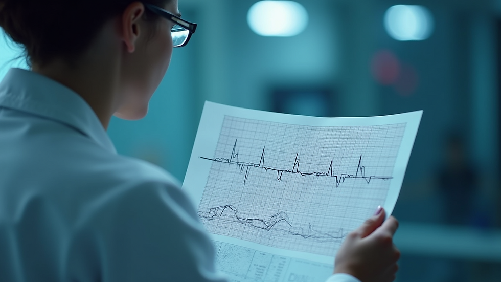
The electrocardiogram (ECG) is a vital tool in modern medicine, providing insights into the electrical activity of the heart. Among the various components of an ECG, the QRS complex is particularly significant, as it represents the depolarization of the ventricles. Understanding the normal and abnormal QRS complexes can help healthcare professionals diagnose a range of cardiac conditions. In this post, we will explore the characteristics of the QRS complex, differentiate between normal and abnormal findings, and review what these abnormalities may indicate.
Understanding the QRS Complex
The QRS complex is a crucial part of the ECG waveform, typically following the P wave and preceding the T wave. It consists of three distinct components: the Q wave, R wave, and S wave.
Q Wave: This is the first negative deflection after the P wave. It is often small and may not be present in all leads.
R Wave: The R wave is the first positive deflection following the Q wave. It is usually the tallest part of the QRS complex.
S Wave: This is the negative deflection that follows the R wave.
The duration and morphology of the QRS complex can provide valuable information about the heart's electrical conduction system.
Normal QRS Complex
A normal QRS complex typically has a duration of 0.06 to 0.10 seconds (60 to 100 milliseconds) and is narrow in appearance. The amplitude can vary, but it is generally well-defined and symmetrical. In a standard 12-lead ECG, the QRS complex should appear consistent across the leads, with slight variations depending on the lead placement. The normal morphology indicates that the electrical impulses are traveling through the ventricles efficiently, suggesting a healthy heart.

Abnormal QRS Complexes
Abnormal QRS complexes can indicate various cardiac issues, ranging from benign conditions to serious pathologies. Here are some common types of abnormal QRS complexes and what they may represent:
1. Wide QRS Complex
A wide QRS complex is defined as one that exceeds 0.10 seconds. This can occur due to several reasons, including:
Bundle Branch Block: This condition occurs when there is a delay in the conduction through one of the bundle branches, leading to a wider QRS complex.
Ventricular Rhythm: In cases of ventricular tachycardia or ventricular fibrillation, the QRS complex may appear wide and bizarre.
Hyperkalemia: Elevated potassium levels can also lead to a widening of the QRS complex.

A Wide QRS Complex on ECG
2. Abnormal QRS Morphology
The morphology of the QRS complex can also provide insights into underlying conditions. Abnormal shapes may include:
Notched QRS: This can indicate left ventricular hypertrophy or other structural heart diseases.

Delta Wave: Seen in Wolff-Parkinson-White syndrome, this indicates an accessory pathway for conduction.

QRS Fragmentation: This may suggest myocardial scar or ischemia.

QRS Complex Fragmentation on ECG
3. Inverted QRS Complex
An inverted QRS complex can be indicative of several conditions, including:
Myocardial Infarction: In certain leads, an inverted QRS may suggest a previous heart attack.
Left Ventricular Hypertrophy: This can also cause changes in the QRS morphology, leading to inversion.

An inverted QRS Complex on ECG
4. QRS Complex with ST Segment Changes
The relationship between the QRS complex and the ST segment is crucial. Abnormalities in the ST segment, such as elevation or depression, can indicate:
Acute Myocardial Infarction: ST elevation in conjunction with a QRS complex can signify an ongoing heart attack.

Ischemia: ST segment depression alongside a normal or abnormal QRS complex may suggest myocardial ischemia.

ST Segment Depression on ECG
Clinical Significance of Abnormal QRS Complexes
Recognizing abnormal QRS complexes is essential for timely diagnosis and treatment. Here are some clinical implications:
Risk Stratification: Patients with wide QRS complexes may be at higher risk for arrhythmias and sudden cardiac death.
Guiding Treatment: Identifying the underlying cause of an abnormal QRS complex can guide treatment decisions, such as the need for medications or interventions like catheter ablation.
Monitoring Progression: Changes in the QRS complex over time can indicate disease progression or response to treatment.
Conclusion
The QRS complex is a vital component of the ECG that provides essential information about the heart's electrical activity. Understanding the differences between normal and abnormal QRS complexes can aid in diagnosing various cardiac conditions. By recognizing the significance of these abnormalities, healthcare professionals can make informed decisions regarding patient care. As you continue to explore the world of ECG interpretation, remember that each QRS complex tells a story about the heart's health. Stay vigilant, and always consider the broader clinical context when evaluating ECG findings.


Register for FREE and get notified every time a new post is added to KIIP by Dr. Saghiv's website. Stay updated all the time with added blog posts about health, wellness, kinesiology, talent acquisition, job seeking, leadership, military service, and more.

Services by Dr. Moran Sciamama Saghiv:
Tags associated with this blog post:
ECG, electrocardiogram, EKG, heart rhythm, cardiac monitoring, heart rate, cardiac test, heart electrical activity, cardiac cycle, P wave, QRS complex, T wave, PR interval, QT interval, ST segment, sinus rhythm, sinus bradycardia, sinus tachycardia, arrhythmia, atrial fibrillation, AFib, atrial flutter, ventricular tachycardia, VT, ventricular fibrillation, VFib, premature beats, PVC, PAC, bundle branch block, BBB, left bundle branch block, LBBB, right bundle branch block, RBBB, heart block, AV block, first degree block, second degree block, third degree block, myocardial infarction, heart attack, STEMI, NSTEMI, ischemia, infarction, repolarization abnormality, depolarization, conduction disturbance, cardiac ischemia, ST elevation, ST depression, T wave inversion, U wave, Q wave, abnormal ECG, normal ECG, 12 lead ECG, ECG leads, lead I, lead II, lead III, aVR, aVL, aVF, precordial leads, V1, V2, V3, V4, V5, V6, limb leads, augmented leads, lead placement, electrode placement, Einthoven triangle, cardiac conduction, SA node, AV node, bundle of His, Purkinje fibers, cardiac axis, axis deviation, left axis deviation, right axis deviation, ECG artifacts, baseline wander, electrical interference, electrode misplacement, ECG interpretation, ECG technician, cardiologist, ECG tracing, ECG printout, ECG waveform, ECG pattern recognition, ECG education, ECG analysis, exercise ECG, stress test, Holter monitor, event monitor, telemetry, portable ECG, wearable ECG, smartwatch ECG, remote cardiac monitoring, ECG abnormalities, cardiac diagnostics, cardiac electrophysiology, clinical ECG, ECG simulator, ECG software, ECG awareness, ECG training, ECG for athletes, ECG in medicine, heart health monitoring.


Comments